May is Melanoma Awareness Month
Did you know that in 2017, approximately 7,200 Canadians were diagnosed with Melanoma? Melanoma is the most dangerous form of skin cancer that has been on the rise in Canada and across the world. If melanoma is diagnosed early, there is a high probability of successful treatment and curability! Staying vigilant and examining all lesions on your body regularly can help you stay on top of our skin health.
Here is a case example of melanoma with Dr. Nakatsui.
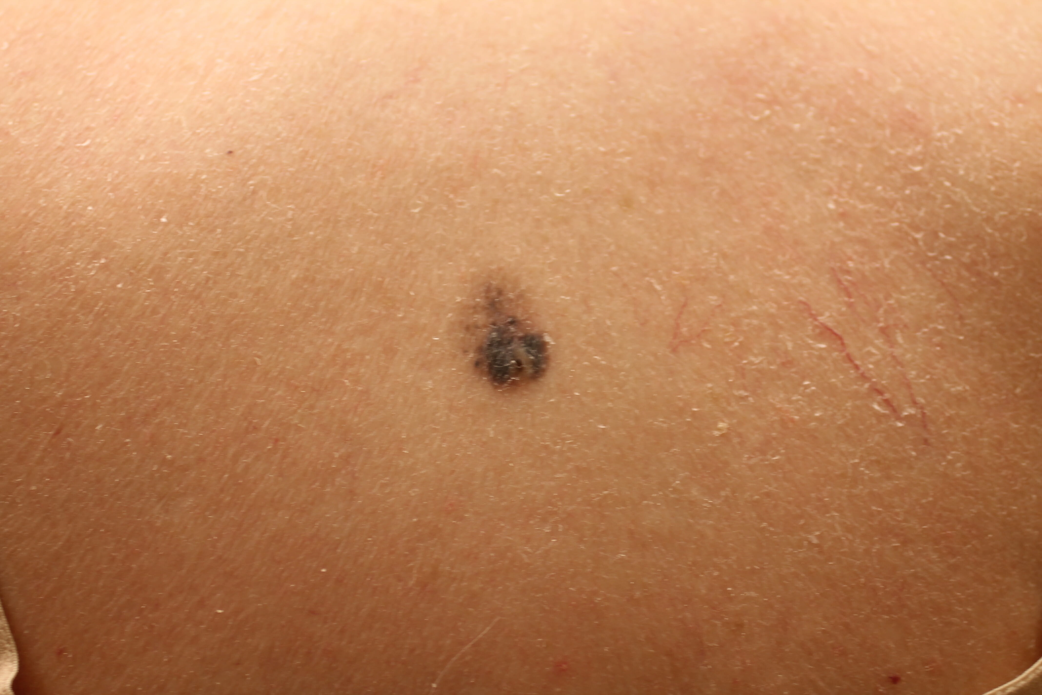
One of Dr. Nakatsui’s patients presented this mole to him and when presented this mole (above). Dr. Nakatsui had a strong suspicion that this oddly shaped mole could be something sinister. Through a diagnostic biopsy performed by Dr. Nakatsui at the Groot DermaSurgery Centre, the results confirmed that the mole was indeed a malignant melanoma, the most worrisome form of skin cancer.
What made Dr. Nakatsui concerned about the mole? We can look at the ABCDE’s of mole checking.
A – Asymmetry: The mole is asymmetrical in shape. If you draw a line through the middle, it is not perfectly symmetrical.
B – Border: The border is uneven and ill-defined. Malignant melanomas typically have uneven, scalloped, or notched borders.
C – Colour: The colour variation in this mole was subtle; however there are elements of light and dark brown, and even some shades of dark blue. Having a multitude of colours in a single mole can be a warning signal. These colours can be reds, browns, blues, whites, and/or blacks.
D – Diameter: The physical size of the mole was another concern for Dr. Nakatsui. The mole was approximately 2.0cm x 1.6cm in size. Malignant melanomas usually are larger than a pencil eras in diameter (6mm), but many malignant melanomas can be smaller than 6mm in size when they are first detected.
E – Evolution: This patient noted that the mole had changed in a relatively short period of time. It grew in size rapidly and it changed in colours within a few months. Any change in colour, shape, elevation, or any other trait, or even new symptoms such as crusting, bleeding, or itching should be examined by your family doctor or dermatologist.
It can be difficult to determine whether a mole is good or bad and the best way to be certain is to have your family doctor or dermatologist examine your moles.
Keep tabs on your moles – Take monthly photos of any or all moles. Use a ruler and place it beside the mole and take a photo. A couple weeks to a month later, take another photo with the ruler again and determine if there is any change to the mole in size, shape, colour, etc. If you do note changes, it is time to see your doctor.
Avoid sun exposure! Wear hats, sunscreen, long shirts to cover your arms, and long pants to cover your legs. Sun exposure can increase the chances of malignant melanomas and also increase the risk of other forms of skin cancers such as basal cell carcinomas and squamous cell carcinomas.
There is a genetic component as well. Ask your immediate and extended family members if they have had melanoma.
When in doubt, check it out!