Birthmarks


Most birthmarks are made of blood vessels bunched together in the skin. They can be flat or raised and pink, red, or blue in color. Ten out of 100 babies have vascular birthmarks. Their cause is unknown. Some will go away on their own, whereas others need to be treated. There are several different types of vascular birthmarks, such as hemangiomas and port-wine stains (also known as a nevus flammeus).
Treatment of vascular birthmarks
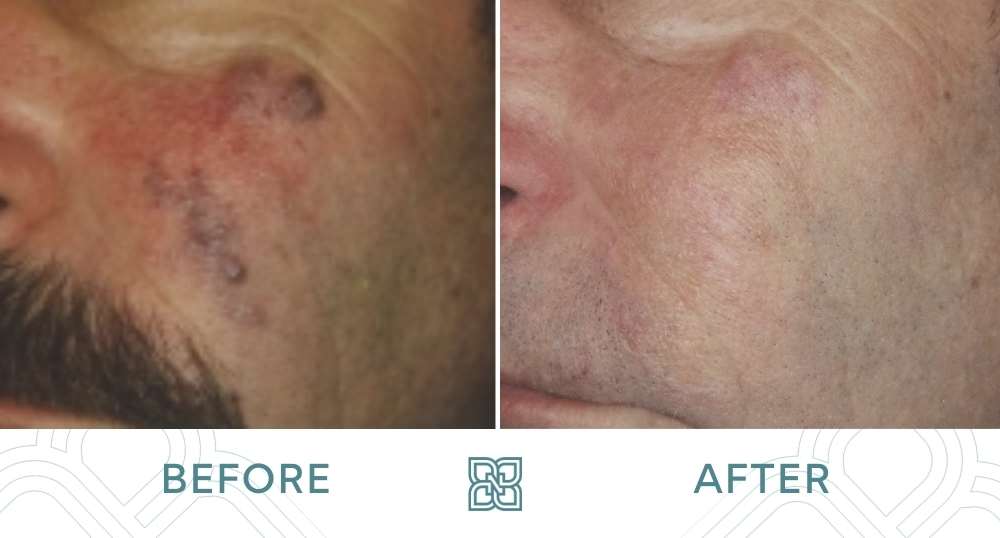
The prescribed treatment will depend on the type of birthmark and in many cases, a combination of lasers are used. Several laser sessions are generally needed for successful treatment with the number of sessions depending on how dark and thick the birthmark is. Fortunately, these types of lesions are typically neither dangerous nor malignant. However, if a hemangioma is close to the eye, glaucoma (high pressure of the eyeball) may occur in very rare cases, and consultation with an ophthalmologist may be necessary. Some vascular birthmarks are small, requiring only a few laser pulses, while others are very large, requiring hundreds to thousands of laser pulses. For these larger ones, we will sometimes treat them under general anesthetic. In a large number of cases (particularly those on the face), treatment of these lesions at Nakatsui DermaSurgery (formerly Groot DermaSurgery Centre) is completely covered by Alberta Health.
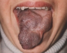
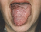
Before and After Vascular Birthmark Photos


**Actual patients. Individual results may vary.
Treatments
Frequently Asked Questions
What is a birthmark?
What Are Vascular Tumors And Malformations?
What Are Strawberry Hemangiomas?
What Are Port Wine Hemangiomas?
A port wine hemangioma is an abnormal collection or network of blood vessels present beneath a layer of otherwise normal skin. The dense network of vessels is the remainder of extra blood tissue that was present during the first month of embryonic life. A port wine hemangioma was so named because the skin appears as though a red, pink or purple liquid such as port wine has been poured over it.
Port wine hemangiomas may be present at birth and grow at the same rate as the normal surrounding skin. In the third, fourth, or fifth decades of life, a port wine hemangioma may become thicker or spongier than the adjacent normal skin and the surface of the hemangioma, which may have been quite smooth during the first decades of life, may develop an irregular and lumpy cobblestone appearance.
What Are Cavernous Hemangiomas?
What Are High Flow Vascular Stains?
High flow vascular stains look very similar to port wine stains but they are associated with increased arterial flow. Upon initial presentation, it can be extremely difficult to distinguish the two conditions. Some features that are more commonly present in high flow vascular stains include heterogeneous stain colour, warmth, peripheral pallor, and sharp, archipelago-like borders.
Unlike port wine stains, these high flow stains are often resistant to laser therapy. Ultrasound and MRI may be helpful in diagnosing high flow vascular stains.
What Treatments Are Available For Vascular Birthmarks?
Many forms of therapy have been used on hemangiomas in the past, however most have been abandoned because they are either ineffective or because they create another deformity that is as undesirable as the hemangioma itself.
Surgeons have removed port wine hemangiomas and reconstructed the area with skin grafts. Such procedures entail a significant amount of surgery, and the scars that result are often quite objectionable. X-rays, which were used in the past, are now known to be potentially dangerous and are no longer used to treat port wine hemangiomas. A variety of agents have been injected into the involved skin but with no significant success. Liquid nitrogen and the carbon dioxide laser have also been used and proven to be ineffective. Tattooing has been attempted on hemangiomas, but the results are temporary, and merely camouflage the birthmark. The best available treatment options may be the use of lasers which target the vasculature in the skin, such as the Vbeam Perfecta Pulsed Dye and Cutera Xeo long pulsed lasers.
How Do The Vascular Removal Lasers Work?
Do you have another question that wasn’t addressed here? Please feel free to contact us with any questions or concerns you may have!


